Most of us likely know someone with breast cancer. After all, it is the most common cancer affecting women in Singapore, with more than 13,000 cases from 2018 to 2022 alone. Thankfully, with advanced screening techniques, we have a better shot at early detection and treatment of breast cancer.
Two of the most common screening tools are mammograms and breast ultrasounds. Both are non-invasive, effective ways to detect abnormalities in breast tissues, though they work differently and serve different purposes depending on your age, breast type, and symptoms.
Not sure which breast screening method is right for you? Let’s compare and learn more about each screening method.
What Is a Mammogram?

A mammogram is a low-dose X-ray imaging tool to detect breast tumours. As the gold standard of breast screening, mammograms can identify early stages of breast cancer. X-rays are a form of radiation absorbed in varying amounts depending on the density of the tissues. The amount of X-rays that pass through the body is processed by a detector, forming images that represent ‘shadows’ of the body structures. Denser structures like tumours and fibrous tissues absorb more X-rays and appear white, while fats and air appear dark grey or black.
Mammograms are especially useful in detecting microcalcifications, a hallmark of early-stage breast cancer. These are calcium deposits that absorb X-rays and show up as bright white specks on ultrasound. Our cells contain calcium ions, which are used for many cellular functions. In the early stages of cancer, as some cancer cells grow rapidly, they might outgrow their blood supply and die. Dying cells release calcium ions and crystallise, forming the distinguishable white specks.
What Is a Breast Ultrasound?
Breast ultrasound uses sound waves that bounce off organs and tissues to return echoes. Ultrasound machines process these echoes to form the typical black-and-white images called sonograms.
Breast ultrasounds are often used as a follow-up to an abnormal mammogram to distinguish between a solid mass and a fluid-filled cyst. Solid structures reflect sound waves better than fluids, creating a pattern of echoes which appear as a lighter grey (hypoechoic) on the image with an irregular shape. In contrast, fluid-filled cysts appear as a dark grey circle (anechoic) with smooth and thin walls.
Ultrasounds are especially useful for women with denser breast tissue, often seen in younger women below 35. A dense breast tissue means a higher proportion of fibrous tissue than fatty tissue. In a mammogram, radiation does not distinguish between fibrous tissue and a solid mass, making it hard to detect tumours in younger women. However, in an ultrasound, dense tissues reflect more sound waves than solid tumours. Denser tissues appear brighter on a sonogram than a solid tumour, making ultrasound a better diagnostic tool for younger women.
Key Differences Between Mammogram and Ultrasound
| Aspect | Mammogram | Ultrasound |
| Technology | Uses low doses of X-rays (ionising radiation). | Uses high-frequency sound waves (non-ionising). |
| Safety | Generally safe, but involves minimal exposure to radiation. | Completely safe, preferred for high-risk individuals like pregnant women. |
| Appearance | Dense breast tissue and tumours both appear white, making them hard to distinguish. | Tumours appear as dark (hypoechoic) areas against a lighter background. |
| Microcalcifications | Highly effective at detecting microcalcifications, an early sign of breast cancer. | Cannot detect microcalcifications. |
| Distinguishing Lumps | Less effective in differentiating solid masses from fluid-filled cysts. | Excellent for distinguishing solid masses from cysts based on their echo patterns and shapes. |
| Common Use | Gold standard for routine breast cancer screening. | Often used as a follow-up to abnormal mammogram results or for younger women with dense breast tissue. |
When Should You Choose One Over the Other?
Choosing between a mammogram and an ultrasound depends on your age, breast tissue type, and symptoms. In many cases, doctors may recommend both to ensure the most accurate diagnosis.
Mammograms are the gold standard for breast cancer screening. Unlike ultrasounds, mammograms are specific for breast cancer. Mammogram screenings are recommended once per year for women aged 40 to 49, and once every two years for those 50 and above.
Ultrasounds are preferred for younger women with denser breast tissue where small tumours may be hidden, or pregnant women who wish to avoid any radiation. They are also used as a follow-up to mammograms, or when a palpable lump is felt but not observed in a mammogram.
What to Expect During Each Procedure?
Both ultrasounds and mammograms are relatively quick procedures, taking about 20 to 30 minutes.
Mammogram
Before the screening, you will be asked some questions regarding your personal details and family history. Let the doctor know if you have breast implants, are breastfeeding, or are pregnant. If you have prior mammograms, be sure to prepare copies of the diagnostic imaging records so that the doctor can compare them with the new ones.
During the mammogram, you’ll be asked to remove any jewellery, undress from the waist up and wear a gown. One breast at a time is placed between two plastic plates that briefly compress the breast to spread out the tissue for a clear X-ray image. This process will be repeated with different angles (top-to-bottom, and side-to-side) to obtain different projections.
The compression might feel uncomfortable or cause mild pressure, but it only lasts a few seconds. Most women find the experience manageable, and the entire procedure usually takes about 20 minutes. It is best to schedule your mammogram after your menstruation when your breasts are not so tender.
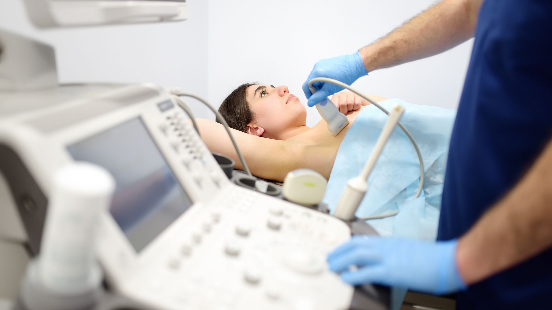
Ultrasound
A breast ultrasound is a painless and non-invasive procedure, typically lasting 15 to 30 minutes. You’ll lie on your back or side, and a technician will apply a small amount of gel to your breast. Then, a handheld device called a transducer will be gently moved over the skin. This device sends sound waves into the tissues and receives the reflected ones, which are transmitted through a machine to form live images. You might feel a slight pressure as the technician moves the probe around, though it should not feel painful. Let your physician know the moment you feel any sharp pain.
Conclusion
Understanding the difference between a breast ultrasound and a mammogram is key to taking charge of your breast health. While mammograms are the gold standard for routine screening and early detection, ultrasounds are valuable for evaluating the type of breast lumps, especially for women with dense breast tissue.
Don’t wait for symptoms to appear. Regular breast screening saves lives by detecting early signs of breast cancer so that you can receive timely treatment. What are you waiting for? Book your breast screening at Centre Screening and Surgery today!
Safeguard Your Health Today
At Centre for Screening and Surgery, we prioritise delivering quality and comfortable early cancer screening and treatment using minimally invasive procedures. If you suspect you might have a breast tumour and are looking for a breast screening, book an appointment with us today!